Index / Work / N° 09
Project N°09 of 24
CategorySimulation
Year2024

Biomedical Image Processing & Cell Area Quantification

Developed an automated image processing pipeline in MATLAB to identify, filter, and quantify the spatial area and centroid locations of SARS-CoV-2 viral particles from transmission electron microscopy (TEM) images.

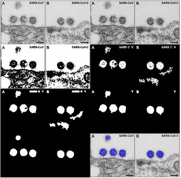
Key Engineering Contributions

  1. 01
    Image Segmentation: Implemented Otsu's method to binarize grayscale TEM images, successfully isolating the viral particles from the background cellular environment.
  2. 02
    Morphological Filtering: Applied custom morphological closing operations and strict geometric thresholds (area boundaries and eccentricity ≤ 0.75) to automatically filter out pixel noise, artifacts, and incomplete cell boundaries.
  3. 03
    Quantitative Data Analysis: Calculated and converted the pixel-based area of the identified cells into physical dimensions, successfully extracting the centroid coordinates and determining a cumulative viral area of 41,151 nm2 across the analyzed sample.

Visual Documentation

Training images - pipeline of image processing the raw TEM images to masking the
Figure 1
01.png
Training images - pipeline of image processing the raw TEM images to masking the SARS-CoV-2 particles and determining their area.
Testing images - pipeline of image processing the raw TEM images to masking the
Figure 2
02.png
Testing images - pipeline of image processing the raw TEM images to masking the SARS-CoV-2 particles and determining their area.